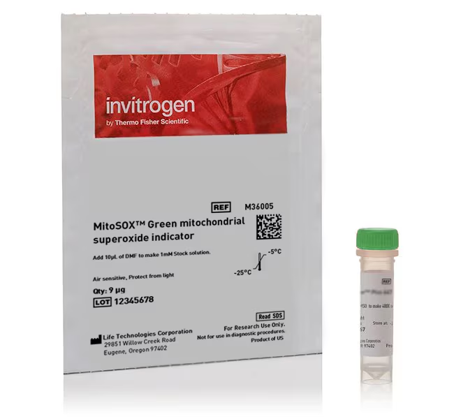
MitoSOX Green and MitoSOX Red superoxide indicators are novel fluorogenic dyes specifically targeted to mitochondria in live cells. Oxidation of the MitoSOX reagent by mitochondrial superoxide produces bright green or red fluorescence.
Features of these indicators include:
• Readily oxidized by superoxide but not by other ROS- or RNS-generating systems
• Use for live cell imaging
• Rapidly and selectively targeted to the mitochondria
• MitoSOX Green indicator: absorption/emission maxima of ∼488/510 nm (traditional FITC/GFP filter set)
• MitoSOX Red indicator: absorption/emission maxima of ∼396/610 nm* (custom filter set recommended)
*Note that two excitation peaks may be observed for MitoSOX Red indicator, as it is excited at both 510 nm and 396 nm. While 510 nm will excite the superoxide oxidation product, it can also excite non-specific products, so we recommend using a 396-nm excitation for more selective detection of mito superoxide when using MitoSOX Red indicator (1).
MitoSOX Green indicator is offered as one vial or a pack of five vials. MitoSOX Red indicator is offered as one vial or a pack of ten vials. A variety pack of one vial each of MitoSOX Red and MitSOX Green indicators is also available.
Consult user Manual for solubility instructions.
Quick, easy detection of mitochondrial superoxide in live cells
The production of superoxide by mitochondria can be visualized in fluorescence microscopy using MitoSOX superoxide indicators. They permeate live cells where they selectively target mitochondria. They are rapidly oxidized by superoxide but not by other reactive oxygen species (ROS) and reactive nitrogen species (RNS). The oxidized product is highly fluorescent.
MitoSOX indicators may be used to distinguish artifacts of isolated mitochondrial preparations from direct measurements of superoxide generated in the mitochondria of live cells. They may also provide a valuable tool in the research of agents that modulate oxidative stress in various pathologies. In addition, these indicators have been used in metabolic flux assays using high-content instruments (2). MitoSOX indicators have also been used to detect mitochondrial superoxide using flow cytometery (3).
References
1: Robinson, Kristine M., et al. Selective fluorescent imaging of superoxide in vivo using ethidium-based probes. Proceedings of the National Academy of Sciences 103.41 (2006): 15038-15043.
2: Little, Andrew Charles, et al. High-content fluorescence imaging with the metabolic flux assay reveals insights into mitochondrial properties and functions. Communications biology 3.1 (2020): 1-10.
3: Kauffman, Megan E. et al. MitoSOX-Based Flow Cytometry for Detecting Mitochondrial ROS. Reactive oxygen species (Apex, N.C.) vol. 2,5 (2016): 361-370.
| Code | Description |
|---|---|
| M36005 | Catalog Number: M36005 Unit Size: 9 µg Indicator: MitoSox Green Color: Green Excitation/Emission: ∼488/510 nm Quantity: 1 Vial |
| M36006 | Catalog Number: M36006 Unit Size: 5 x 9 µg Indicator: MitoSox Green Color: Green Excitation/Emission: ∼488/510 nm Quantity: 5 Vials |
| M36008 | Catalog Number: M36008 Unit Size: 10 x 50 µg Indicator: MitoSox Red Color: Red Excitation/Emission: ∼396/610 nm Quantity: 10 Vials |
| M36007 | Catalog Number: M36007 Unit Size: 50 µg Indicator: MitoSox Red Color: Red Excitation/Emission: ∼396/610 nm Quantity: 1 Vial |
| M36009 | Catalog Number: M36009 Unit Size: 2 vials Indicator: MitoSox Green, MitoSox Red Color: Green, Red Excitation/Emission: 488/510 nm (green), 396/610 nm (red) Quantity: 2 Vials (1 Green, 1 Red) |

