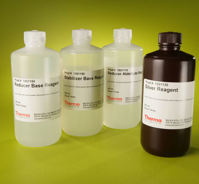
Thermo Scientific Pierce Color Silver Stain Kit detects proteins electrophoresed in 1D and 2D polyacrylamide gels using metallic silver to produce color variations that facilitate differentiation of protein bands and spots.
Features of the Color Silver Stain Kit:
• Customizable—kit is a reagent set that allows customized formulation of working reagents and staining protocols for particular gel formats and silver-staining applications
• Sensitive—detects many proteins at 0.1ng per square millimeter in standard-thickness gels
• Broad protein detectability—stains a wider range of protein types than monochromatic silver stains (proteins that do not bind silver are revealed as yellow bands or spots)
• Color differentiation—gel-separated proteins and polypeptide-containing macromolecules stain in five basic colors: black, blue, brown, red and yellow
• Profiling resolution—multicolored detection of DNA, lipids and polysaccharides in addition to typical proteins provides additional landmarks to aid in 2D gel profiling
The Color Silver Stain Kit contains four reagents and a flexible protocol for staining proteins in 1D and 2D electrophoresis gels. The method requires more time than monochromatic silver stains but provides greater flexibility in adapting the concentration of working solutions to obtain effective staining with different gel types and thicknesses and unusual protein targets. Proteins stain as black, blue, brown, red or yellow bands or spots according to a combination of their specific charge and other characteristics. Once results are known for a particular protein of interest, these slight color differences provide for easy band and spot identification.
Includes:
• Four reagent solutions (500 mL each): silver, reducer base, reducer aldehyde and stabilizer base
The Color Silver Stain Kit enables highly sensitive and versatile staining of proteins electrophoresed in 1D and 2D polyacrylamide gels. Generally, bands containing less than 1ng of protein are detectable with this silver stain. Proteins stain in reproducibly distinct colors visible against an amber background. Nucleic acids, as in the products of PCR, that have been electrophoresed in polyacrylamide gels also may be stained using this kit.
Several slightly different silver staining methods were developed in the early 1980s. All methods depend on the kinetics of impregnation, reduction of silver ions to elemental silver and its complexation to protein nucleation sites in a porous gel matrix. The Color Silver Stain Kit uses a weakly acidic solution of silver nitrate for gel impregnation, followed by development in formaldehyde at alkaline pH. After incubation in the Stabilizer Base Working Solution, silver nitrate and formaldehyde are either reacted or diffused from the gel.
When a stained gel is viewed over a bright white light source, several distinct colors may be recognized. Five basic colors commonly visible in protein samples with the Color Silver Stain are black, blue, brown, red and yellow. Color differences correspond to differences in protein amino acid composition, although detailed predictions are not possible. Nevertheless, color patterns are reproducible for a given sample and staining time and temperature, and they may be used to distinguish overlapping proteins and other subtleties in 2D systems.
Regardless of gel size and thickness, the Color Silver Stain Kit is well characterized and supported by this complete procedure for obtaining excellent staining results for routine and special electrophoresis experiments involving polyacrylamide gels.
| Code | Description |
|---|---|
| 24597 | Catalog Number: 24597 |

